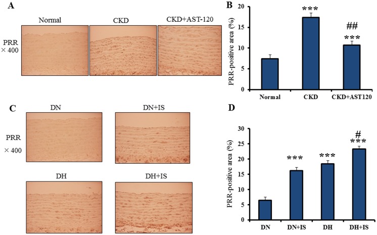
Figure 1. Immunohistochemistry of PRR in rat aorta.
A. Immonohistochemical localization of PRR in the aortas of normal, CKD and AST-120-treated CKD rats. B. Quantitative data of PRR in the aortas of normal (n = 9), CKD (n = 8) and AST-120-treated CKD rats (n = 8) (mean±SE). ***p<0.001 vs normal, ##p<0.001 vs CKD. C. Immonohistochemical localization of PRR in the aortas of DN, DN+IS, DH and DH+IS rats. D. Quantitative data of PRR-positive area in the aorta of DN, DN+IS, DH and DH+IS rats (mean±SE, n = 8). ***p<0.001vs DN, #p<0.05 vs DH.

