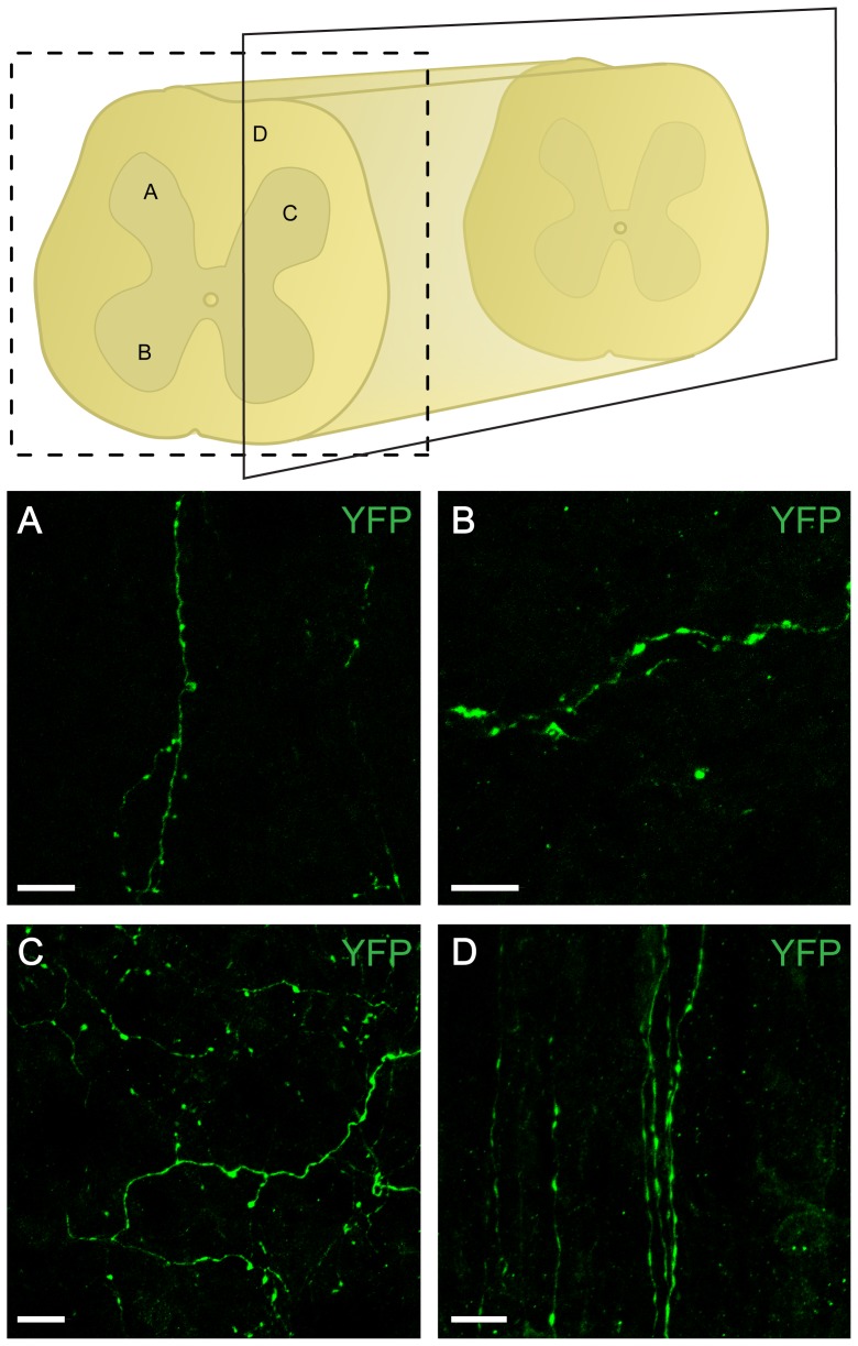
Figure 4. YFP positive fibers in the lumbar spinal cord originating from A11.
Schematic of fiber localization in the lumbar spinal cord. Representative micrographs of YFP-labeled fibers in transverse (A, B) and parasagittal (C, D) lumbar spinal cord sections. Fibers were found in the dorsal (A) and ventral horn (B) as well as in the grey (C) and the white matter (D). Scale bar: 10 µm.

