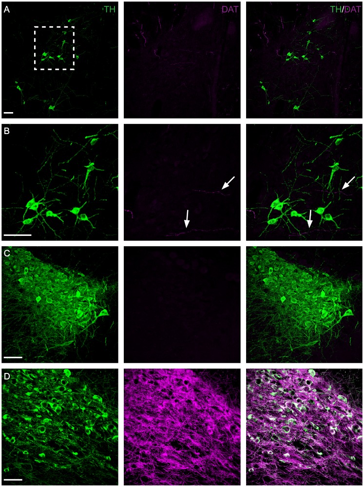
Figure 7. Absence of dopamine transporter (DAT) expression in A11.
Immunohistochemistry targeted against TH (green) and DAT (magenta) (A–D). Representative micrographs showing the absence of DAT labeling in the middle region of A11 (A, B). Note the DAT positive but TH negative fibers in A11 (B) (arrow). (C) As expected DAT is absent in the locus coeruleus (LC) which served as a negative control. Representative micrograph of TH and DAT labeled neurons and fibers in the ventral tegmental area (VTA) (D). Note the absence of DAT expression in the A11 and the LC in contrast with the intense expression in the VTA (positive control). B: Higher magnification of boxed area in A. Scale bar: 50 µm.

