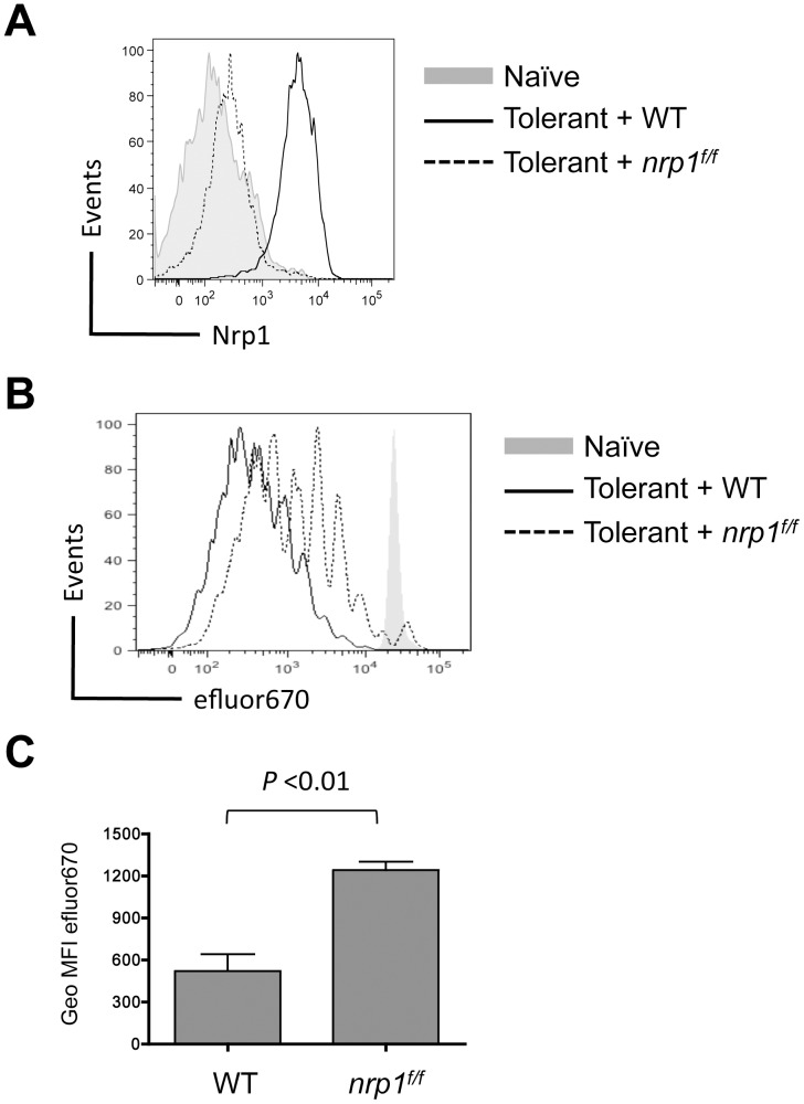
Figure 2. Nrp1 expression corresponds with a modest increase in CD8+ T cell proliferation in response to self-antigen.
Naive WT or Nrp1-deficient CD8+ T cells were labeled with efluor670 cytoplasmic dye and co-transferred into B6 (naive) or Alb∶Gag (tolerant) mice. (A) Nrp1 expression on WT and nrp1f/f T cells 3 days after transfer is compared in the overlaid histograms. (B) Dilution of efluor670 dye in transferred T cells was assessed 3 days after transfer and is displayed in overlayed histograms. (C) Geometric mean fluorescent intensity of efluor670 in T cells from either WT or Nrp1-deficient T cells (lower dye expression corresponds to more proliferation) is pooled from 4 separate experiments, each with 3 mice per group. Error bars are standard error of the mean (SEM) with P value indicated.

