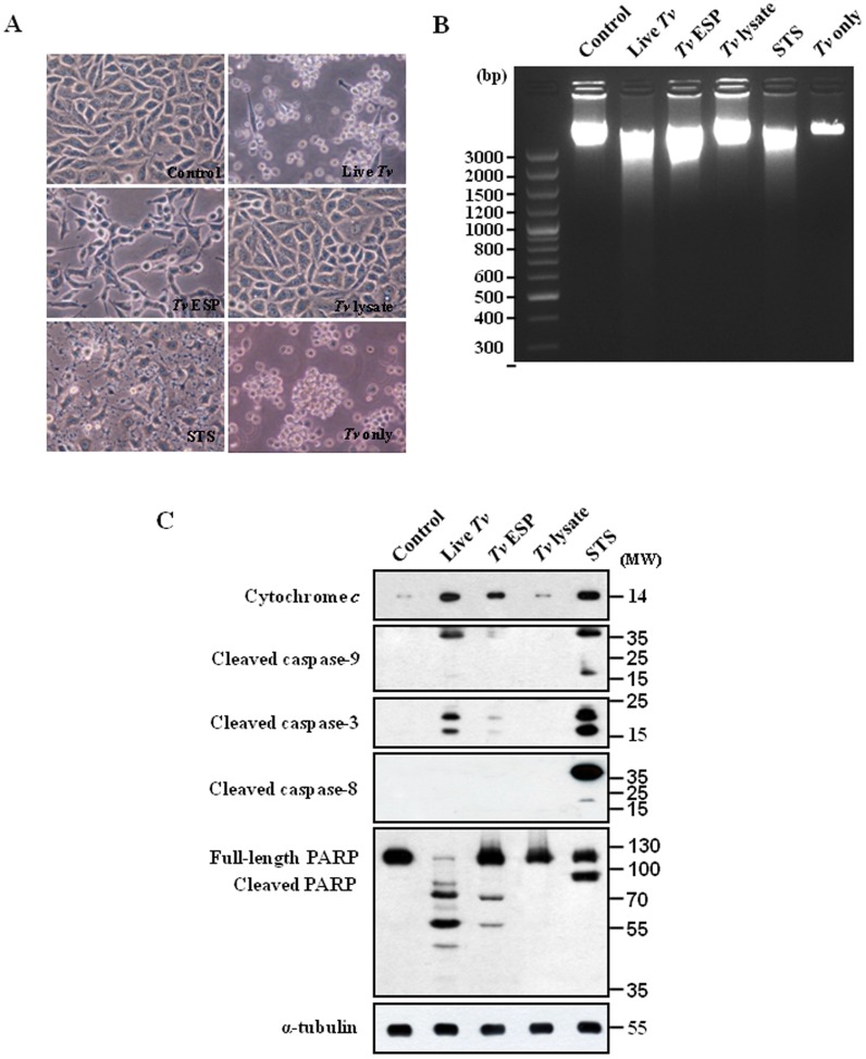
Figure 1. Mitochondria-dependent apoptosis in SiHa cells after treatment with Trichomonas vaginalis antigens.
(A) Micrographs of SiHa cells incubated with live T. vaginalis (MOI = 2), T. vaginalis excretory and secretory products (ESP) (100 µg/mL), T. vaginalis lysate (100 µg/mL), or STS (1 µM) for 16 h. (B) DNA fragmentations of the SiHa cells were determined by agarose-gel electrophoresis. (C) The cytosolic fraction (only for dectection of cytochrome c) or protein extracts (detection for the others beside of cytochrome c) of the SiHa cells were subjected to western blotting using the indicated antibodies. A representative image from three independent replicates is shown.

