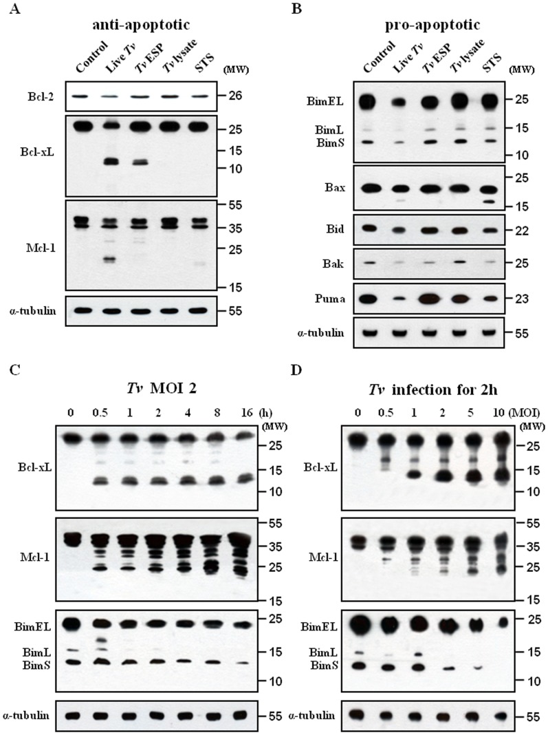
Figure 3. Western blot analysis of Bcl-2 family proteins in SiHa cells treated various T. vaginalis antigens.
SiHa cells were treated as in Fig. 1A, the protein extracts were analysed with (A) anti-Bcl-2, anti-Bcl-xL, anti-Mcl-1, and anti-α tubulin antibodies or (B) anti-Bim, anti-Bax, anti-Bid, anti-Bak, and anti-Puma antibodies. SiHa cells were stimulated with T. vaginalis (MOI = 2) for the indicated times (C) or at the indicated MOI for 2 h (D). The protein extracts were analyzed by western blotting using anti-Bcl-xL, anti-Mcl-1 and anti-Bim antibodies. A representative result of three independent replicates is shown.

