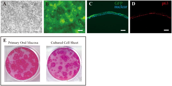
Figure 2. Successful generation of transplantable epithelial cell sheets from GFP rats.
(A) Phase-contrast image of oral mucosal epithelial cells. (B) Fluorescence microscopy showing GFP-positive cells from a donor GFP rat. (C) Cross-section of a GFP-positive cell sheet. (D) Basal cells of cultured epithelial cell sheets express p63. (E) Both primary oral mucosa and cultured cell sheets contained sufficient numbers of progenitor cells. Scale bars = 200 µm (A, B) and 50 µm (C, D).

