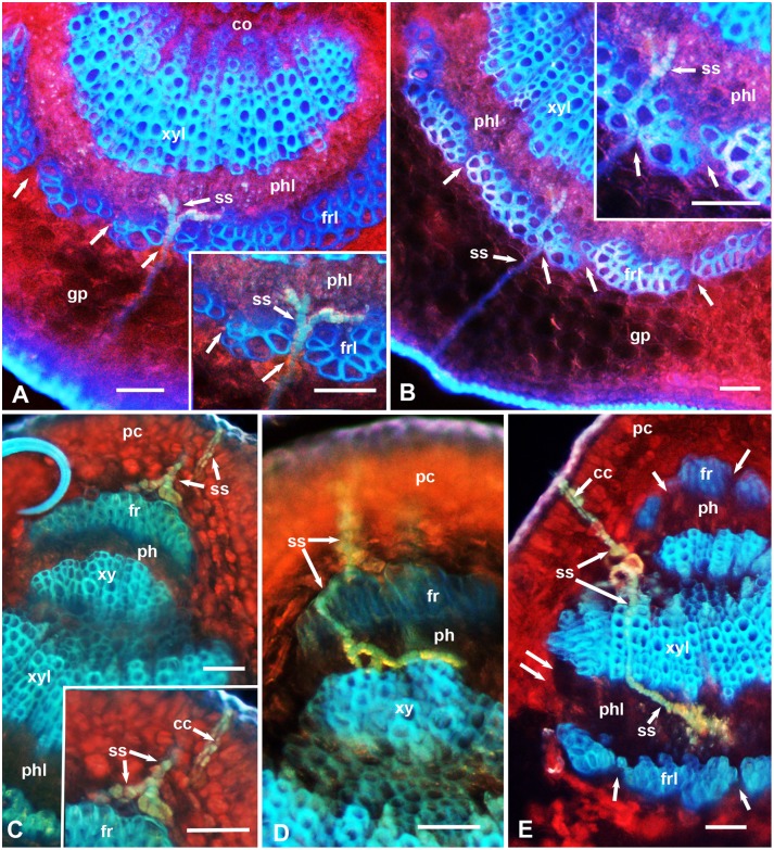
Figure 2. Epifluorescence micrographs showing stylet sheaths (ss) of D. citri in cross sections of the midrib in leaves of the relatively resistant (UN-3881) plants.
In A–E, the upper (adaxial) leaf side is up, and the lower (abaxial) side is down; unlabeled single arrows indicate smaller gaps in the fibrous ring; double arrows indicate wider gaps in this ring at the sides of the vascular bundle. Abbreviations: cc, central canal in the stylet sheath; co, core cells; fr, upper fibrous ring; frl, lower fibrous ring; gp, ground/spongy parenchyma; pc, palisade parenchyma cells; ph, upper phloem; phl, lower phloem; ss, stylet sheath; xy, upper xylem; xyl, lower xylem. Scale bars = 50 µm.

