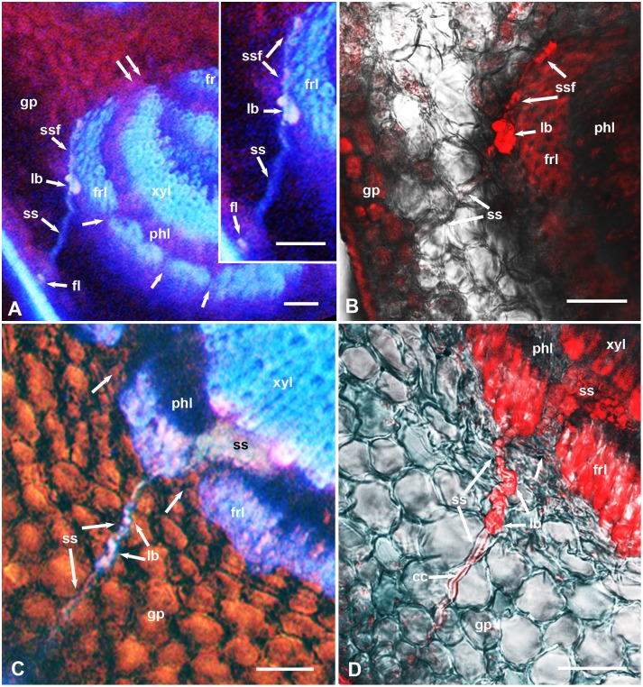
Figure 4. Stylet sheath interactions with the ground parenchyma and fibrous ring in cross sections, stained with propidium iodide, in the midrib of D. citri-resistant Poncirus trifoliata (A & B) or the relatively susceptible xCitroncirus (C & D) plants.
A & C, epifluorescence microscopy images; B & D, confocal laser scanning images of the same or adjacent sections as those in A and C, respectively; differential interference contrast was also used in B–D to show cell boundaries. Unlabeled single arrows indicate smaller gaps in the fibrous ring; double arrows indicate wider gaps in this ring at the sides of the vascular bundle. Abbreviations: cc, central canal inside sheath; fl, stylet sheath flange; fr, upper fibrous ring; frl, lower fibrous ring; gp, ground/spongy parenchyma; lb, larger blebs (secretion bursts) in the stylet sheath; pc, palisade parenchyma cells; phl, lower phloem; ss, stylet sheath; ssf, part of stylet sheath going around fibrous ring; xyl, lower xylem. Scale bars = 50 µm.

