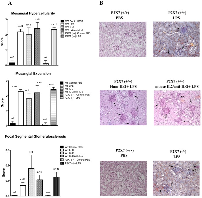
Figure 2. Histology features.
Classical histology (Ematoxylin Eosin, PAS) of kidney biopsies was evaluated after several times from LPS infusion (24hrs, 72 hrs and 7 days). Three main features were observed: 1-mesangial hypercellularity, 2-mesangial expansion and 3-focal segmental glomerulosclerosis that were determined in a semi-quantitative basis (score 0–3 for the former two parameters, score 0–0.3 for glomerulosclerosis). Relevant results observed after 7 days are presented in this figure (A). Specific patterns are shown in (B); stains are hematoxylin eosin for all with the exception of Masson Blue stain for LPS alone.

