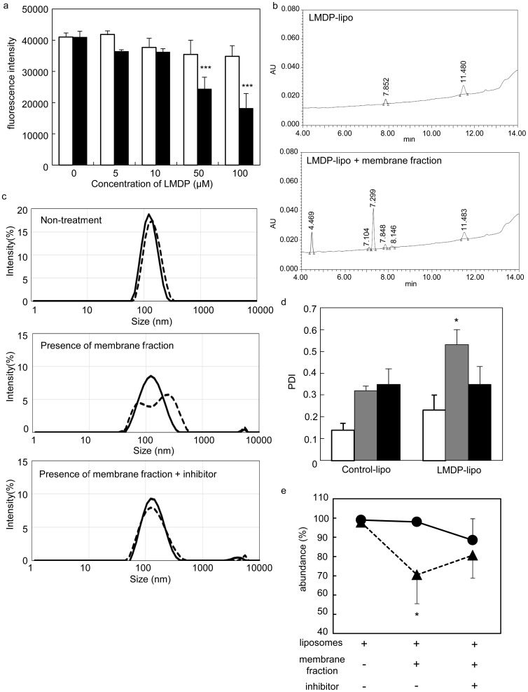
Figure 4. Characterization of LMDP-lipo cleavage by γ-secretase.
(a) Competitive inhibition of LMDP in liposomes. The fluorescence peptide probe was incubated with Control-lipo (EPC/DOPE/CHEMS) or LMDP-lipo (EPC/DOPE/CHEMS/3 mol% LMDP) at the indicated LMDP concentrations in the presence of the membrane fraction of A549 cells at 37°C overnight. White and black columns indicate Control-lipo or LMDP-lipo, respectively. (b) Representative chromatograms of LMDP-liposomes with or without the membrane fraction as determined by HPLC analysis from three independent experiments. (c) Size distribution of liposomes in the presence of the membrane fraction obtained from A549 cells with or without γ-secretase inhibitor was measured using the Zetasizer nano. Solid line and dotted line indicate Control-lipo and LMDP-lipo, respectively. (d) Polydispersity index (PDI) of the peak using Control-lipo or LMDP-lipo. White, gray and black columns indicate liposomes alone, liposomes in the presence of an A549 cell membrane fraction or liposomes in the presence of a membrane fraction with 10 µM DAPT, respectively. (e) Abundance distribution of the peak for Control-lipo (black circles) and LMDP-lipo (black triangles) under the same conditions as (d). (Values and bars represent the means and SD, respectively. *, P<0.05 and ***, P<0.001 versus 0 µM LMDP (a), Control-lipo (c, d), n = 3.

