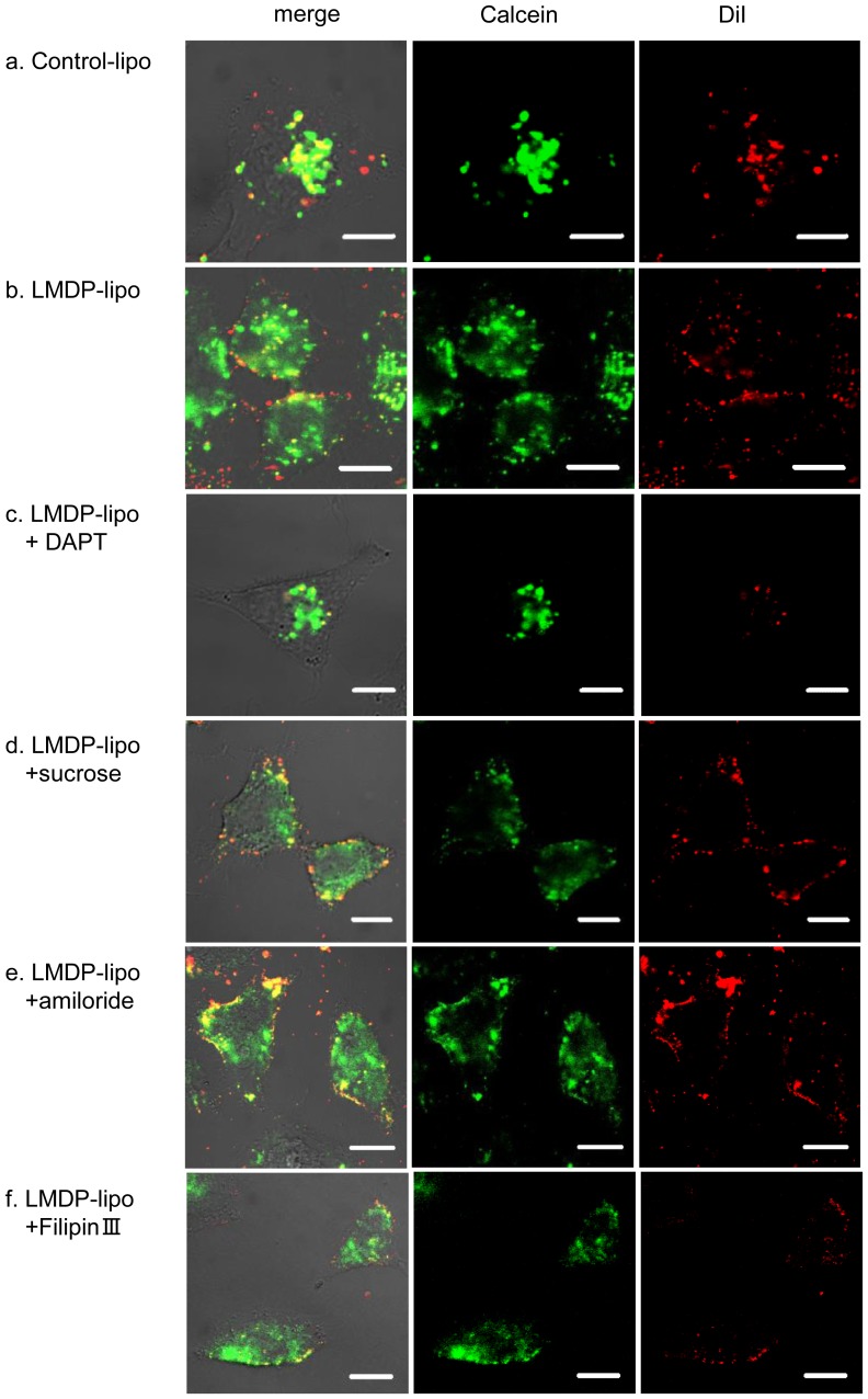
Figure 6. Calcein release from LMDP-lipo into cells.
DiI-labeled and calcein encapsulated Control-lipo (EPC/DOPE/CHEMS) (a), LMDP-lipo (EPC/DOPE/CHEMS/5 mol% LMDP) (b), LMDP-lipo in the presence of 50 µM DAPT (c), LMDP-lipo in the presence of 0.4 M sucrose (d), LMDP-lipo in the presence of 2.5 mM amiloride (e) and LMDP-lipo in the presence of 5 µg/mL FilipinIII (f) were added to A549 cells and incubated at 37°C for 1 h. The intracellular location of CM-DiI (red) and calcein (green) was observed by CLSM. Red signal indicates liposomes. Scale bars, 10 µm.

