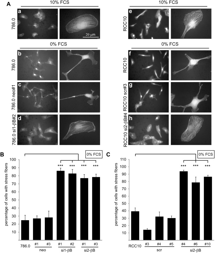
Figure 2. Knockdown of Activin B stabilizes actin stress fibers in ccRCC cells.
(A) Subconfluent parental 786.0 and RCC10 cells, control clones (786.0 neo#1, RCC10 scr#3) and Activin B knockdown clones (786.0 si1-βB#2, RCC10 si2-βB#4), respectively, were maintained in the presence of 10% FCS or serum starved (0% FCS) for 3 hours. Micrographs show TRITC-labeled Phalloidin staining of the actin cytoskeleton. (B) and (C) Quantification of the indicated 786.0 (B) and RCC10 cells (C) with actin stress fibers upon serum starvation for 3 hours. Phalloidin stained cells were classified by microscopic analysis and at least 200 cells were counted per experiment. Bars represent the mean of four independent experiments, error bars indicate standard deviation. Statistical significance was determined by ANOVA analysis and denoted by asterisks: ***P<0.001.

