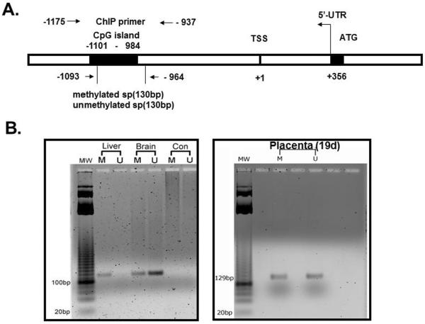
Figure 1.
A. Schematic representation of the putative CpG island in relation to the transcriptional start site (TSS) within the 5' flanking region of the glut3 gene. Location of primers employed in the methylation specific PCR (MSP) and chromatin immunoprecipitation (ChIP) associated PCR are represented by arrows. B. Representative gel demonstrating the glut3 amplification product (~130 bp) obtained by employed either methylated or unmethylated set of forward and reverse primers on genomic DNA obtained from liver (does not express Glut3), brain (expresses high amounts of Glut3) and placenta (expressed lesser amounts of Glut3 compared to brain). CON = negative PCR control.

