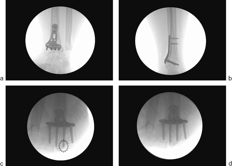
Fig. 3.

Intraoperative fluoroscopic control of a osteosynthesis of distal radius fracture by a volar plate (case No. 59). (a) AP view. The most distal ulnar screw does not cross the DRUJ. (b) Lateral view. No screws appear to cross the dorsal cortical bone of the distal radius. (c) Skyline view. The second distal radial screw crosses the dorsal cortical bone of the distal radius. (d) Skyline view. After change of screws, the second distal radial screw does not cross the dorsal cortical bone of the distal radius.
