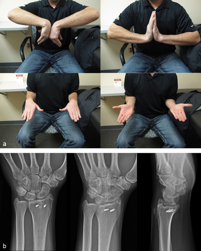
Fig. 3.

(a) Clinical photographs show flexion, extension, pronation, and supination of the injured wrist (left) compared with the contralateral uninjured wrist (right) at 1 year postinjury. (b) PA, oblique, and lateral radiographs of the left wrist at 1 year postinjury show a healed radial styloid fracture and maintained reduction of the radiocarpal joint without dorsal or ulnar translation of the carpus.
