
Figure 4.
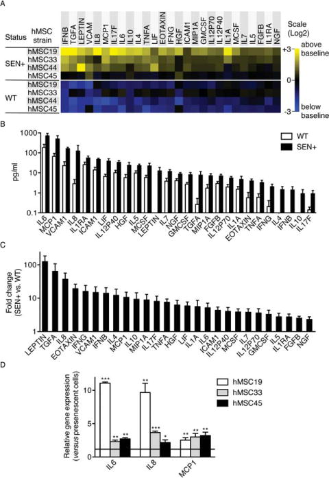
Senescent hMSCs secrete higher levels of inflammatory mediators compared to WT cells. (A): Soluble factors secreted by the indicated cells were detected by antibody arrays and analyzed as described in the text, Materials and Methods, and Supporting Information. WT, wild-type primary hMSCs; SEN+, gamma-irradiated hMSCs. For each protein, all signals were averaged and used as the baseline. Signals higher than baseline are displayed in yellow; signals below baseline are displayed in blue. The numbers in the heat map key (right) indicate log 2-fold changes from the baseline. Only proteins significantly oversecreted in SEN+ cells (p < .05; compared with WT cells) are represented. (B): Levels of proteins (normalized to 105 cells per milliliter) significantly oversecreted in SEN+ cells compared with WT cells. (C): Fold changes in the levels of proteins significantly oversecreted in SEN+ cells compared with WT cells. When protein levels where under the detection limit, a baseline of 0.1 pg/ml was considered. (D): Relative expression levels of the major SASP components IL6, IL8, and MCP1 were quantified by real-time RT-PCR in three different isolates of presenescent and replicative-senescent hMSCs. α-Tubulin was used as endogenous control. *, p < .05; **, p < .01; ***, p < .005, versus controls. Error bars represent SEM (n = 3). Abbreviations: hMSC, human mesenchymal stem cell; WT, wild type.