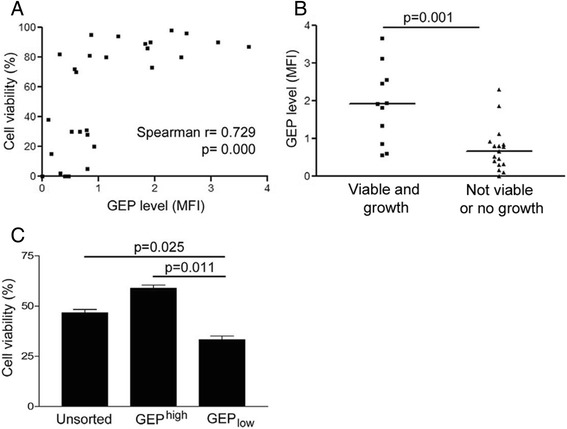
Figure 2.

Correlation of GEP expression with success rate of HCC primary culture. (A) GEP levels significantly correlated with the viability of the cells freshly isolated from HCC tissues (n = 28, Spearman’s ρ correlation coefficient = 0.729, p = 0.000). GEP level was measured by flow cytometry and expressed as mean fluorescence intensity (MFI) after subtracting the non-specific background signal (isotype control). (B) GEP levels (MFI) of the cells with viability > 70% and could grew on culture plate successfully (viable and growth) were significantly higher than those failed or viability <70% (not viable or no growth) (n = 28, p = 0.001). (C) Hep3B cells were sorted for surface GEP expression by magnetic sorting. Unsorted control cells, sorted GEPhigh and GEPlow cells were then cultured in 10% FBS-supplemented AMEM in ultra-low attachment plate for 12 h. Cells were harvested and stained for annexin V-propidium iodide for apoptosis. Cells negative for both annexin V and propidium iodide were defined as viable cells, which were quantified by flow cytometric analysis. Viability of GEPlow cells was significantly lower than unsorted and sorted GEPhigh cells.
