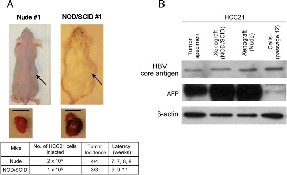
Figure 7.

Tumorigenicity of HCC21 cells in immunocompromised mice. (A) Nude mice and NOD/SCID mice injected subcutaneously with 2x10⁶ and 1x10⁶ HCC21 cells, respectively, after 12 weeks. The middle panel shows the subcutaneous tumors derived from HCC21 cells 12 weeks after injection (scale bar = 10 mm). The lower panel shows the tumorgenicity of HCC21 cells. (B) Protein expression of HBV core antigen and AFP in patient’s tumor specimen, HCC21 xenograft tumors in NOD/SCID and nude mice, and cultured HCC21 cells at passage 12.
