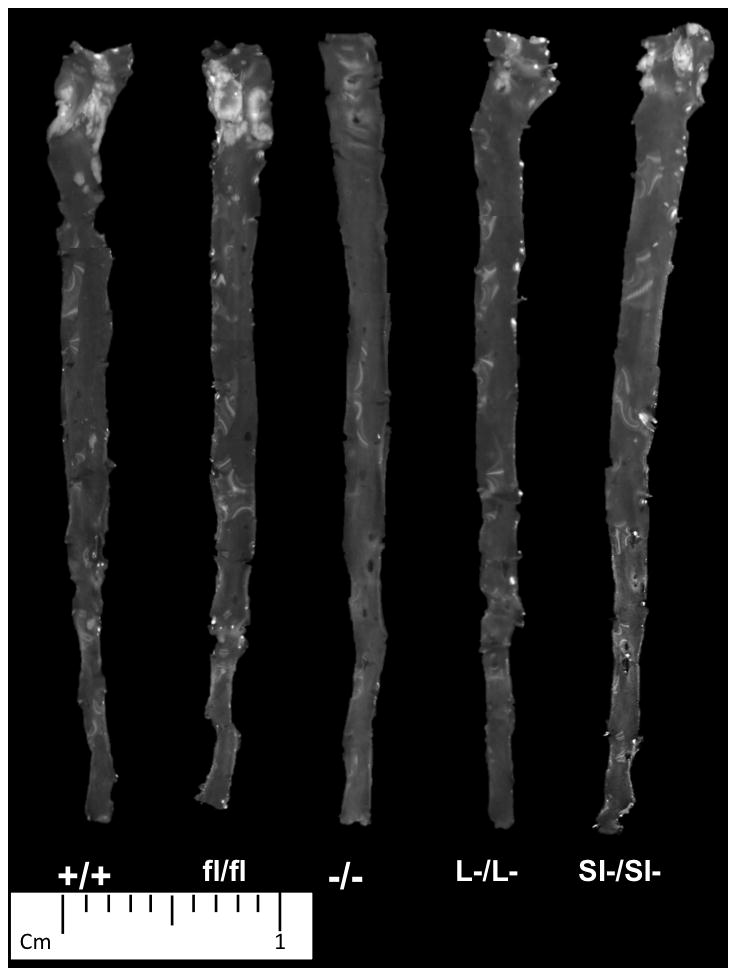
Figure 4. Images of aorta isolated from all SOAT2 knockouts.

At the time of necropsy, the entire length of aorta was excised. Atherosclerotic lesions were noticeable as opaque areas mainly in the areas of ascending aorta and aortic arch. Representative image of each genotype was presented. N = 10 to 12 per genotype.
