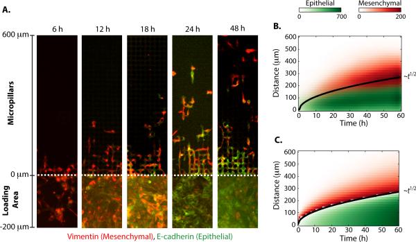
Fig. 3. The dynamics of individual scattering from a collectively migrating front can be understood as a dispersion phenomenon from a moving interface.
(A) Immunofluorescent staining of the pillar region (0 < y) reveals individually migrating mesenchymal cells detaching from a collectively migrating epithelial front. Cells in the rear loading region (y < 0) exhibit phenotypic plasticity and undergo a mesenchymal to epithelial transition (MET) over 24 h. (B) The measured spatial distributions of individual and collectively migrating cells are plotted as a function of time, showing an interface that propagates outward as the square root of time. (C) A solidification model for binary mixtures shows quantitative agreement with experimental data.

