
Fig. 4.
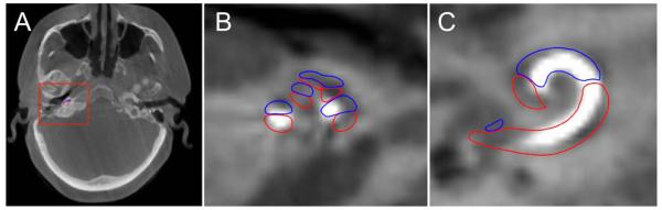
Computed tomography of a right temporal bone following cochlear implant electrode insertion. (A) Axial, (B) magnified oblique axial, and (C) magnified oblique coronal views demonstrating the implant beginning in the scala tympani (outlined in blue) and crossing over into the scala vestibuli (outlined in red) at approximately 180°.