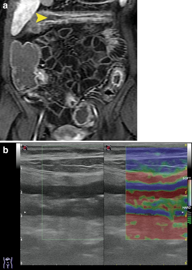
Fig. 10.

Same patient as Fig. 9. a Contrast enhanced MRI: the transverse colon presents localized enhancement mainly in the tunica mucosa and submucosa, arrowhead (suggesting that the disease is in the acute phase [9] ), there is luminal distension of the ascending colon due to partial occlusion of the proximal transverse colon. b Strain elastography of the proximal transverse colon: the appearance of the colonic wall suggests concomitant fibrosis/thickening of the muscular layer (predominantly blue) and inflammation of the mucosa and submucosa (green and red)
