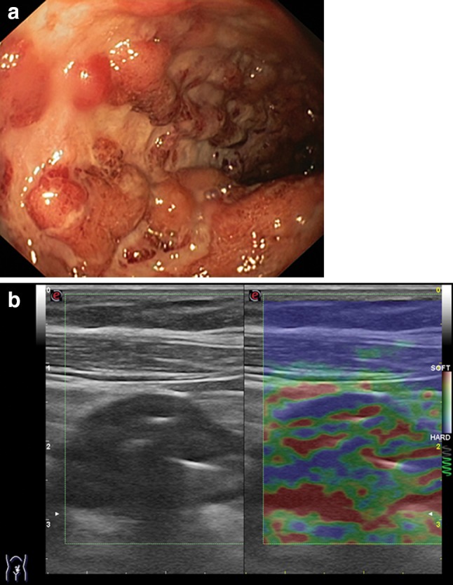
Fig. 8.

a Endoscopy shows the sigmoid colon with areas of congested mucosa separated by deep ulcerations (Mayo endoscopic score 3). b Corresponding strain elastography image of the sigmoid colon showing moderate wall thickening and abnormal elastographic appearance, classified 2BAR (mix of colors: green > blue > red) compatible with the endoscopic image showing very severe disease
