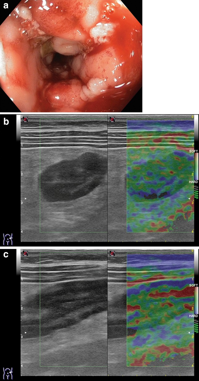
Fig. 9.

a Endoscopy shows the sigmoid colon with extensive mucosal defects, marked edema, cobblestone appearance, bleeding on probing. b, c Strain elastography image of the sigmoid colon classified 2BAR (green > blue with small yellow and red spots) and the distal descending colon classified 2BRA (green > red > blue) these strain elastography patterns correlate with the acute phase of the disease as evidenced by endoscopy
