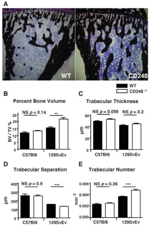
Figure 3. CD248-deficient mice have increased trabecular bone volume.
A, The tibiae of wild-type (WT) and CD248−/− (129SvEv strain) mice were stained with von Kossa’s stain to detect trabecular bone formation (dark brown; counterstained with hematoxylin [purple]). B–E, Micro–computed tomography was performed to measure the trabecular bone structure in WT and CD248−/− mice on the C57BL/6 and 129SvEv genetic backgrounds, assessed as the percentage of bone volume/tissue volume (BV/TV) (B), trabecular thickness (C), trabecular separation (D), and trabecular number (E). Bars show the mean ± SD of 6 samples per group. ** = P < 0.01; *** = P < 0.001, by Student’s t-test. NS = not significant.

