
Figure 4.
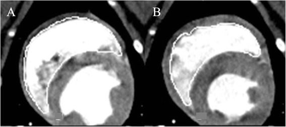
Midventricular short-axis of the right ventricle in cardiac computed tomography (CCT) of an anaesthetised healthy beagle. In CCT-short-axis the endocardial border is manually traced in (A) end-diastolic and (B) end-systolic volume and as in all performed methods the papillary muscles and trabeculae were included in the volumes.