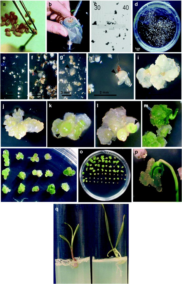
Fig. 1.

Tissue culture based on fragmented mature embryos: methodology, morphogenic pathway of the initiated calli, and shoot regeneration. Aseptic embryo isolation with a lancet under a stereo microscope (a), grinding by extrusion through a sterile nylon mesh, using the rounded tip of the handle of a lancet (b), the resulting mature caryopses reduced to fragments: tissue fragments laid down on micrometric cover glass (c) and in suspension in the liquid basal medium (in a 9-cm Petri dish) before induction in in vitro culture (d). Scheduling of tissue culture within the time scale of 1 week (e–h) and 2–8 weeks (i–p). 2,4-D cell proliferation induction in explants after 1, 4, and 7 days of culture in darkness, respectively (e–g/h). Numerous actively proliferating specimens visible after 4 days (f) doubled their volume within 3 days (g); some of these 7-day-old structures, densely formed with actively dividing cells, appeared as compact and were considered morphogenic (h). Nodular appearance calli visible 2 weeks after the 2,4-D induction (i). Clusters of yellowish-white nodular structures (j), chlorophyll synthesis (large green pigmented areas within the nodular structures, k), development of organs (green tissue rising up to the surface, l), leaf emergence and expansion (m–o), and plantlet regeneration (p) after the calli were transferred to a medium without growth regulators and exposed to light. Healthy plantlets and root development on the rooting medium (q: plantlet on the left starting to develop roots; plantlet on the right showing slightly more developed roots, with lateral growth)
