Figure 5.
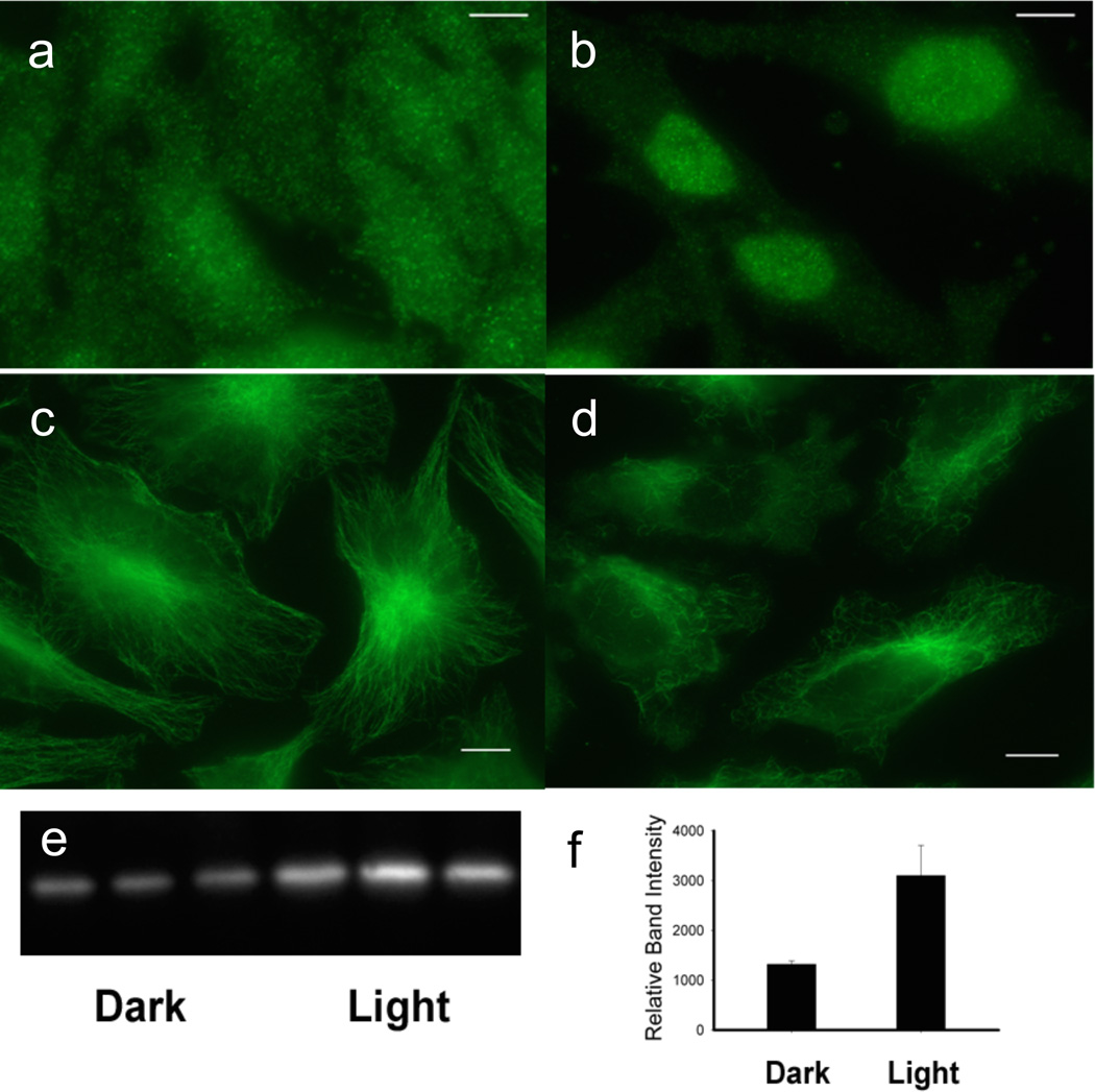
Release of anti-inflammatory agents from erythrocytes and their affect on HeLa cells. (a) – (b) DEX release from C18-Cbl-DEX/C18-Cy5-loaded erythrocytes at 660 nm triggers HeLa cell GRα nuclear localization, where (a) is dark and (b) 660 nm. HeLa GRα visualized with Alexa488 antiRabbit/anti-GRα. (c) – (d) COL release from Cbl-COL/C18-DY800 erythrocytes at 780 nm initiates HeLa microtubule depolymerization, where (c) is dark and (d) 780 nm. HeLa microtubules visualized with Alexa488 antiMouse/anti-tubulin. Bar = 5 µm. (e) – (f) MTX release from C18-Cbl-MTX/C18-Cy7-loaded erythrocytes at 725 nm shifts the thermal stability of DHFR where left 3 lanes (dark) and right 3 lanes (725 nm). GAPDH used as loading control (SI Figure S30).

