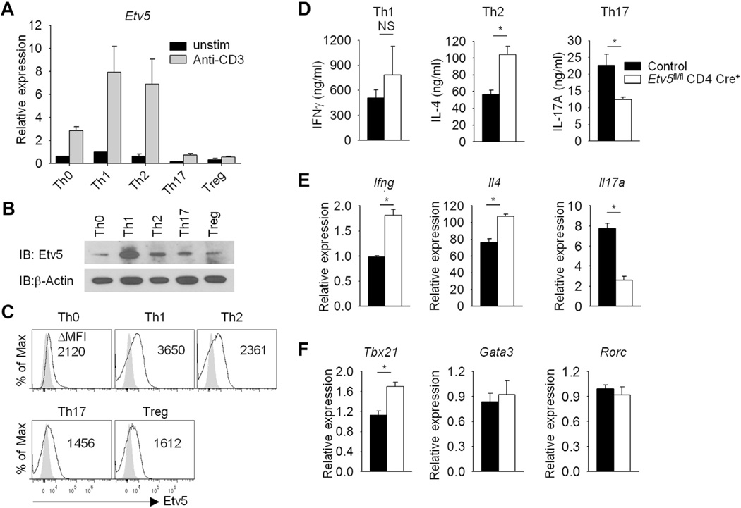
FIG 2.
Helper T-cell differentiation in the absence of Etv5 in T cells. Naive control and Etv5-deficient CD4+CD62L+ T cells were activated (TH0) or cultured under TH1, TH2, TH17, and Treg cell polarizing conditions. Etv5 expression was measured in helper T-cell subsets by using qRT-PCR before and after 6 hours of anti-CD3 stimulation (A), immunoblotting (B), or intracellular staining (C) before anti-CD3 stimulation. Δ Mean fluorescence intensity was calculated by subtracting the background from the signal of Etv5 antibody. TH1, TH2, and TH17 cells were used for assessing cytokine production by means of ELISA after 24 hours of anti-CD3 stimulation (D) and gene expression analysis after (Ifng, Il4, and Il17a; E) or before (Tbx21, Gata3, and Rorc; F) 6 hours of anti-CD3 stimulation by means of qRT-PCR. Data are means ± SEMs of 4 independent experiments (Fig 2, D-F) or means ± SDs of replicate samples (Fig 2, A) and representative of 3 independent experiments with similar results (Fig 2, A-C). *P < .05. NS, Not significant.

