Fig. 3.
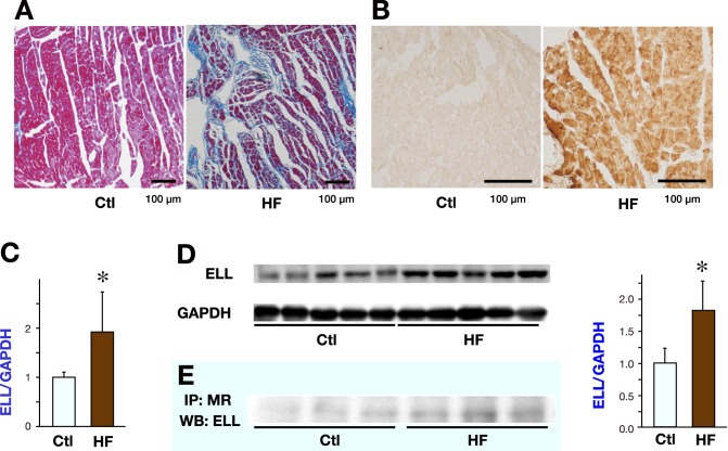
LV fibrosis, ROS generation, ELL expression and ELL-MR interaction in the LVs of 22 weeks old Dahl salt-sensitive rats.
Representative photomicrographs of Azan Mallory staining to evaluate LV fibrosis (A) and representative immunohistochemical staining for 4-hydroxy-2-nonenal to evaluate ROS generation (B) in the LV of a Dahl-salt sensitive rat in the Ctl group and the HF group.
C: RT-qPCR analysis of the mRNA levels of ELL in the LVs of Dahl salt-sensitive rats (n = 8 for each group).
D: WB analysis of the protein levels of ELL in the LVs of Dahl salt-sensitive rats. The images (left) and the results of densitometry (right) of WB analysis (n = 5 for each group).
Data are expressed as mean values ± SD. The Ctl group was arbitrarily set to 1.
*P < 0.05 versus the Ctl group.
E: LV lysate of Dahl salt-sensitive rats was subjected to IP with anti-MR antibody. Immunoprecipitates were subsequently analyzed by Western blotting with anti-ELL antibody.
Ctl, control; ELL, elongation factor eleven-nineteen lysine-rich leukemia; HF, heart failure; IP, immunoprecipitation; LV, left ventricle; MR, mineralocorticoid receptor; ROS, reactive oxygen species; WB, Western blot.

