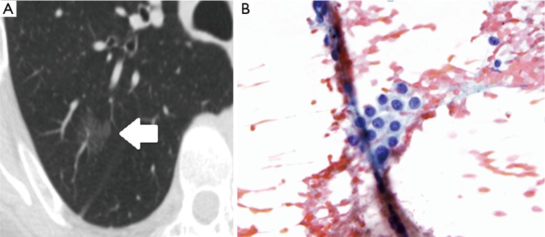
Figure 2.

Radiologic-pathologic correlation for a preinvasive lesion in the adenocarcinoma spectrum. On CT (A), the lesion consists of a purely ground-glass opacity (white arrow), through which vascular structures can still be distinguished. Atypical cells are seen on pathology (B). Given its ground-glass appearance and size of between 5 mm and 3 cm as seen on CT, this lesion likely represents adenocarcinoma in situ, although this diagnosis can only be confirmed after no invasive component is demonstrated upon examination of a fully resected surgical specimen. (B, magnification 40×). CT, computed tomography.
