
Figure 3.
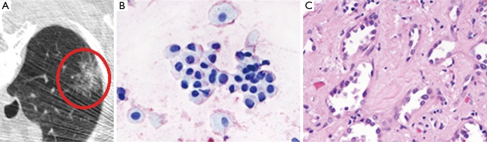
Radiologic-pathologic correlation for an invasive adenocarcinoma. CT evaluation (A) of the right lung shows a subsolid nodule at the periphery, which is greater than 5 mm in size, with a partial solid component (circled). Cytology (B) from the lesion demonstrates atypical bronchioloalveolar cells, with focal stromal invasion demonstrated on surgical biopsy specimen (C), confirming an invasive, T4N0 adenocarcinoma. (B, magnification 40×; C, magnification 20×). CT, computed tomography.