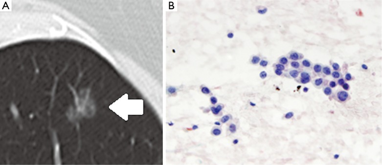
Figure 4.

Radiologic-pathologic correlation for intralesional lucency within a subsolid nodule. On CT (A), a partially ground-glass, partially solid nodule, suspicious for invasive adenocarcinoma, also demonstrates a subtle pseudocavitation at its posterior aspect (arrow), which is associated with a favorable prognosis. Cytology (B) from the specimen demonstrates atypical bronchioloalveolar cells. (B, magnification 40×). CT, computed tomography.
