Fig. 2.
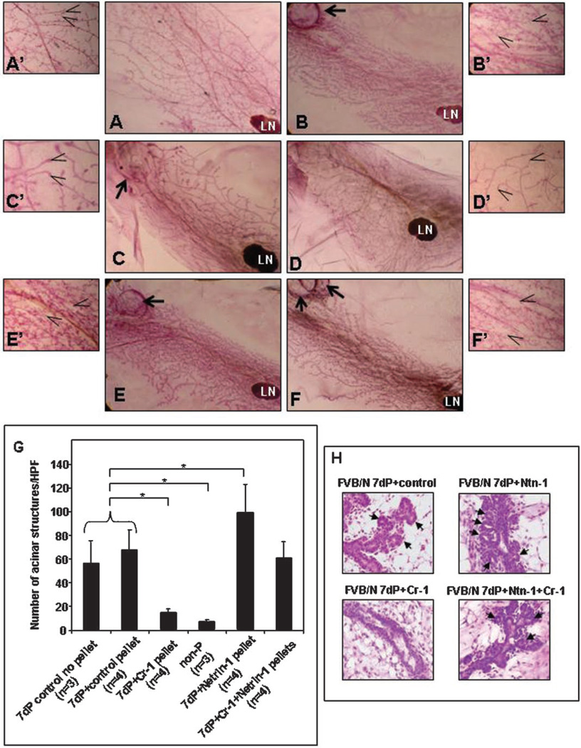
Whole mount (WM) morphology of mammary glands from FVB/N mice. Untreated mammary gland from a 7-day pregnant (dP) FVB/N mouse or with control cholesterol pellet show comparable WM morphology (A,A′ vs. B,B′) and alveolar counts (G). WM morphology of a 7 dP FVB/N mouse containing rmCR-1 releasing pellet (C,C′) shows significantly reduced (P < 0.05) alveolar counts (G) which is similar to the WM morphology (D,D′) and alveolar counts (G) of normal non-pregnant (non-P) pubescent FVB/N control mice. Treatment with rmNetrin-1 caused enhanced development (E,E′) and significant increase (P < 0.05) in alveolar counts (G) in mammary glands of 7 dP FVB/N mice. WM morphology (F,F′) and alveolar counts (G) was similar to that of control mice when both rmCR-1 and rmNetrin-1 pellets were combined. H&E sections in (H) show the histological details of the alveolar structures (arrows) that formed during the different treatment conditions. Arrows in A to F point to surgically implanted pellets. Open arrows in A′ to F′ point to alveolar structures counted for quantification in (G). (LN = lymph node; *P < 0.05). Magnification of A to F = 10×; magnification of A′ to F′ and H = 40×.

