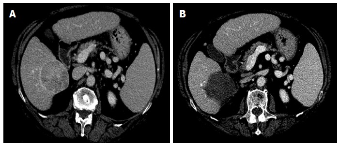
Figure 1.

Representative case of complete ablation of large nodule with multi-fibre technique. A: Computed tomography (CT) scan before laser ablation (LA) session shows a nodular lesion 6 cm in maximum diameter (hepatocellular carcinoma moderately differentiated) localized in the S6 with exophytic growth (exophytic component > 40%); B: CT scan performed 4 wk after LA procedure shows complete necrosis of the tumor. Four illuminations were performed using the pullback technique and the treatment lasted 24 min. The procedure was well tolerated and the patient was discharged from the hospital 24 h after the procedure. The only side effects were mild pain and self-limiting fever lasting for 7 d.
