
Figure 5.
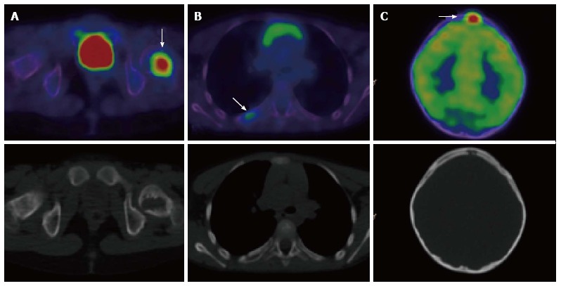
A 7-year-old male with Langerhans cell histiocytosis. Skeletal survey demonstrated an isolated left femoral lesion, confirmed on PET/CT (A, arrow). Additional lesions (arrows) in the right 6th rib posteriorly (B) and in the skull (C) were also identified on the PET/CT scan. Top panel: fused PET/CT images, bottom panel: low dose CT component of the scan. PET/CT: Positron emission tomography/computed tomography.