Figure 6.

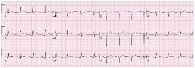
Diffuse ST elevation secondary to acute pericarditis. There is typical depression of the PR segment (seen mainly in leads II and aVF. There is ST elevation in the inferolateral leads with ST depression in lead aVR.

Diffuse ST elevation secondary to acute pericarditis. There is typical depression of the PR segment (seen mainly in leads II and aVF. There is ST elevation in the inferolateral leads with ST depression in lead aVR.