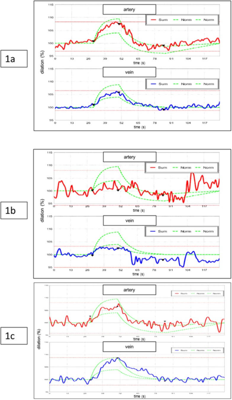
Fig. (1).
(a) Typical behaviour of retinal vessels on flicker stimulation in a healthy eye as measured by Dynamic Vessel Analysis (red: artery; blue: vein). The first 30 seconds show uninfluenced baseline data. The average of uninfluenced vessel diameter measurements is set 100 %. Higher values indicate dilation and lower values contraction as compared to the baseline vessel diameters. At the following flicker light provocation vessel diameters dilate. After flicker stimulation they decrease and may even slightly fall below values at the starting level. After recovery they reach baseline values again. ‘Sum’ is giving all measurements of one patient in their temporal sequence. The area between the two ‘Norm’ curves represents the normal range. (b, c) Typical response of retinal vessels on flicker stimulation in a glaucomatous eye (group 1) as measured by Dynamic Vessel Analysis; (b) before trabeculectomy, (c) after trabeculectomy. A marked improvement of the vessel reaction can be seen.

