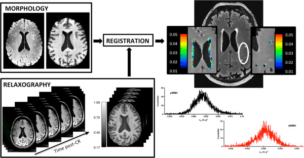
Figure 1.

Data workflow: (Left, top) FLAIR images were linearly registered to high-resolution MPRAGE space using rigid body registration. (Left, lower) Co-registered R1 maps were prepared in high-resolution MPRAGE space after alignment of all variable-TI MPRAGE images to the TI 1800 image collected at the temporal mid-point of CR washout (see Methods for additional details). (Right) Fractional blood volume (vb) maps of pWMHs and dWMHs and corresponding FLAIR image. Insets show a magnified view of the WMHs (outlined in white) with associated color scales. The vb histograms of each WMH are also shown.
