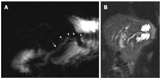
Figure 16.

Mild chronic pancreatitis. A: Coronal oblique thick-slab magnetic cholangiopancreatogram image; B: Coronal single-shot turbo spin-echo T2-weighted (HASTE) image with fat-suppression. There is uniform dilatation of the pancreatic duct (arrow) and prominence of the pancreatic duct side-branches (arrowheads), without significant diffuse thinning of the pancreatic parenchyma (A, B) in keeping with mild chronic pancreatitis.
