
Figure 1.
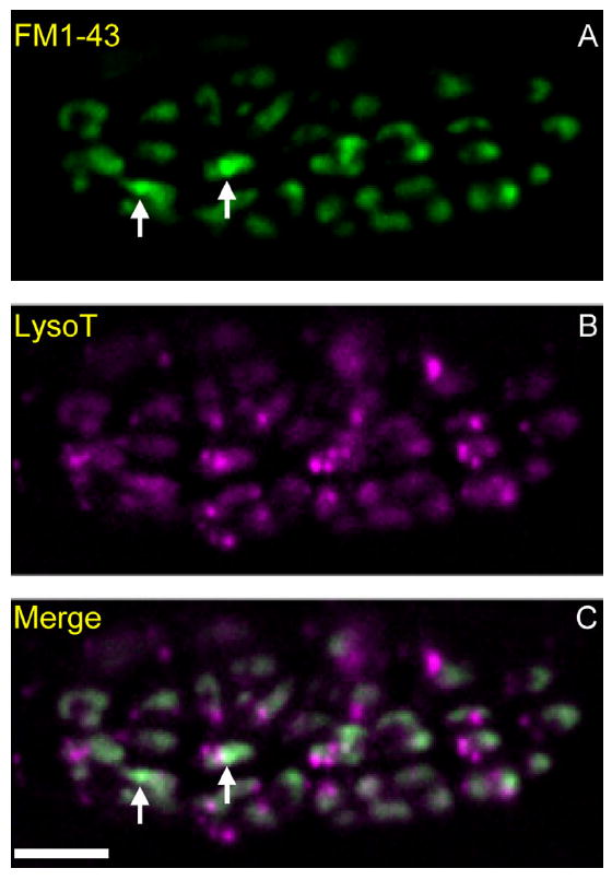
Macroendosomes (MEs) and acidic endosomes (AEs) in a snake motor nerve terminal. A–C: Preparations were incubated with LysoTracker (B; magenta), rinsed, and then electrically stimulated with bath-applied FM1-43 (A; green; C is merged). Punctate acidic endosomes (AEs, magenta in B and C) are visible throughout the terminal. Two macroendosomes (MEs, green) are marked with arrows in A and C. Background FM1-43 “haze” (green) obscures MEs and is due to recently endocytosed 50-nm vesicles that also contain FM1-43. Scale bar = 10 μm in C (applies to A–C).