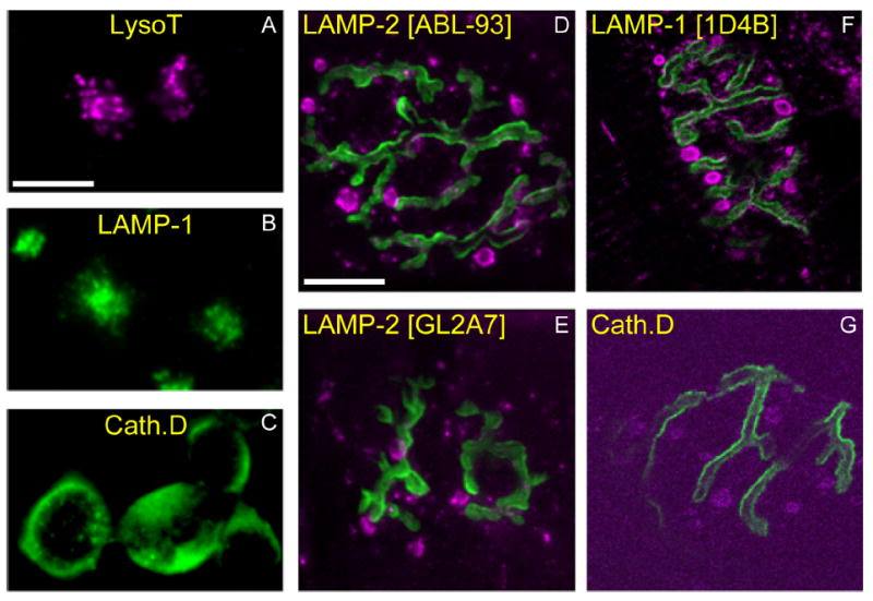
Figure 12.

AEs contain lysosomal marker proteins. A–C: Mouse neuroblastoma (N2a) cells were incubated vitally with LysoTracker (A; magenta), or fixed and immunostained with an antibody against LAMP-1 (1D4B) (B; green) or Cathepsin D (C; green). As expected, small (300–500-nm) vesicles (lysosomes) are seen concentrated near the nuclear compartment. D–G: Mouse muscles (gastrocnemius) were incubated with 488–Bungarotoxin (green) to identify AChRs apposing motor terminals and fixed. Immunostaining with antibodies against various lysosomal proteins (magenta; specific antibodies indicated at top of each panel) revealed a pattern similar to LysoTracker staining of AEs (see Fig. 11F), including large structures outside the terminal. Scale bar = 10 μm in A (applies to A–C) and D (applies to D–G).
