
Figure 5.
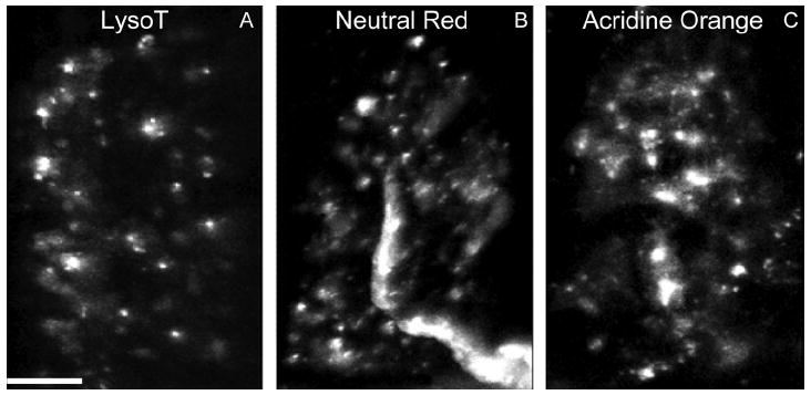
AEs are recognized by conventional supravital acidophilic probes. A–C: Living snake nerve–muscle preparations were incubated with Neutral Red (B) or Acridine Orange (C; both 1 μg/ml, 30 minutes), followed by washing with reptilian saline and imaging as in Figure 4. Characteristics of the small puncta (white) stained by the dyes are similar to LysoTracker-positive structures (AEs) (compare with A; see also Fig. 1B). Scale bar = 10 μm in A (applies to A–C).