Figure 7.

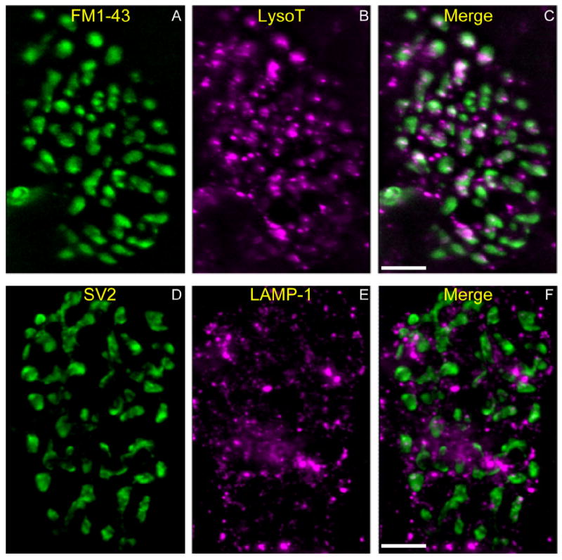
LAMP-1–positive structures in fixed snake terminals resemble living AEs. A–C: Living snake terminal labeled with FM1-43 (A; green) by KCl depolarization (60 mM, 3 minutes) to reveal location of bouton vesicular compartment. AEs are labeled with LysoTracker (B; magenta). Merged image (C) indicates that most AEs are within boutons but not within the vesicular compartments. Note AEs in axon (entering at the lower left). The axon (green) is labeled by FM1-43 because of the dye’s affinity to myelin membrane. D–F: Another snake terminal fixed and immunostained with the synaptic vesicle marker SV2 (D; green) to reveal vesicular compartments. The terminal is also stained with anti-LAMP-1 (Sigma), a marker for lysosomes (E; magenta; F is merged). Note the similar size and number of LysoTracker and LAMP-1–positive vesicles (compare B and E), and the similar distributions of LAMP-1 and LysoTracker mainly outside the vesicular compartment (compare C and F). Scale bar = 10 μm in C (applies to A–C) and F (applies to D–F).
