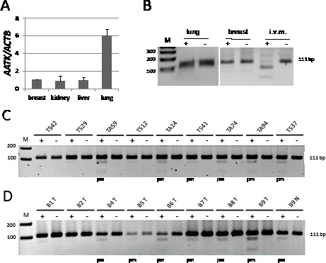
Fig. 2. Expression and methylation of AATK in normal tissues and primary tumors.

A. Expression of AATK was analyzed in normal breast, kidney, liver and lung tissues by quantitative RT-PCR and normalized to ACTB levels (breast=1). B. Methylation of AATK was analyzed in normal lung and breast tissues and in vitro methylated DNA by COBRA. Mock digest (−) and TaqI digest (+) are shown. C. Methylation analysis is shown for primary lung cancer samples (TA=adenocarcinoma, TS=squamous cell carcinoma). D. Methylation analysis in primary breast cancer (T=tumor, N=corresponding normal tissue). Products were resolved on a 2% gel with a 100 bp marker (M). (pm=partially methylated).
