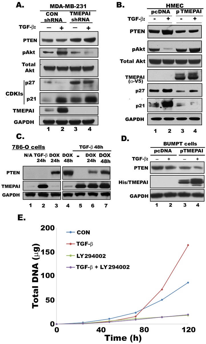
Fig. 5. Effect of TMEPAI on PTEN, Akt, pAkt and CDKIs expression.
A. Relative expression determined by Western blotting of PTEN, phosphorylated Akt (pAkt), total Akt, p21, p27, TMEPAI and GAPDH in MDA-MB-231 cells expressing control (CON shRNA) or TMEPAI shRNA without and with TGF-β (2 ng/ml) treatment. B) Western blots of HMEC expressing control vector (pcDNA) or human TMEPAI (pTMEPAI) in the absence or presence of TGF-β. C) Relative levels of PTEN and TMEPAI in PTEN null 786-O human renal carcinoma cells expressing doxycycline (DOX) inducible PTEN vector in the absence or presence of TGF-β (2 ng/ml) and/or Doxycycline (1μg/ml). D) Relative expression of PTEN protein in mouse proximal tubule cells (BUMPT) expressing pcDNA or mouse TMEPAI (pTMEPAI) without or with TGF-β (2 ng/ml). TMEPAI is detected with anti-His antibody. E) Growth curves of MDA-MB-231 cells in the absence or presence of TGF-β (2 ng/ml) and LY294002(10μM) alone or together.

