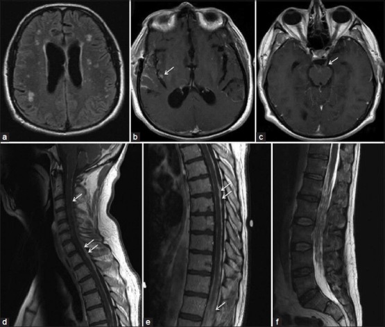
Figure 1.

Brain MRI (a–c) showing diffuse areas of FLAIR signal changes (a) and T1W post-gadolinium sequence with diffuse leptomeningeal and nodular parenchymal enhancement (b, arrow) and cranial nerve enhancement (c, arrow showing CNIII). Post-contrast scan of cervical (d) and thoracic (e) spine showing diffuse nodular leptomeningeal enhancement. This was most significant at the lower lumbar region with almost complete obliteration of normal CSF signal on T2WI (f)
