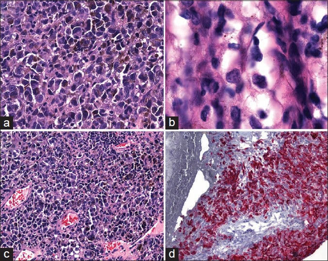
Figure 2.

Hematoxylin and eosin staining of the cauda equina demonstrating: (a, ×100) pleomorphic spindle cells with prominent nucleoli, mitotic figures (b, ×1250), and melanin pigment. Spreading of neoplastic cells along subpial and perivascular spaces (c, ×100). Immunohistochemical stains for melanoma cocktail including Melan-A demonstrate cytoplasmic reactivity (d)
