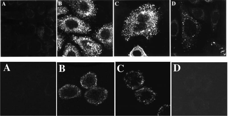
Figure 3. HER2 and transferrin receptor phage antibodies and scFv are internalized into cells.
Top panel: Confocal microscopy with detection of phage antibodies using anti-pVIII antibody. A. Control phage antibody, binds botulinum neurotoxin. B. F5 anti-HER2 phage antibody selected for cellular endocytosis. C. Anti-transferrin phage antibody selected for cellular endocytosis. D. C6.5 anti-ErbB2 phage antibody selected on recombinant ErbB2 [61].
Bottom panel: Confocal microscopy with detection of scFv using an antibody to a Cterminal peptide tag on the scFv. A. Control scFv, binds botulinum neurotoxin. B. F5 scFv selected for cellular endocytosis. C. Anti-transferrin scFv H7 selected for cellular endocytosis. D. C6.5 anti-ErbB2 scFv selected on recombinant ErbB2 [61]. This figure cited images from reference #33.

